Right Shoulder Anatomy Diagram : Shoulder Anatomy | New York, NY | HandSport Surgery Institute / The disk has a great variation in size and shape and eventually undergoes rapid degeneration until it is.
Right Shoulder Anatomy Diagram : Shoulder Anatomy | New York, NY | HandSport Surgery Institute / The disk has a great variation in size and shape and eventually undergoes rapid degeneration until it is.. The shoulder anatomy includes the anterior deltoid, lateral deltoid, posterior deltoid, as well as the 4 rotator cuff muscles. 2.1 bones of the shoulder girdle. The shoulder joint is formed where the humerus (upper arm bone) fits into the scapula. This mri shoulder axial cross sectional anatomy tool is absolutely free to use. This set is often saved in the same folder as.
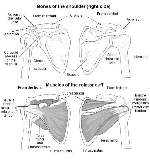
The shoulder muscles consist of the deltoids and the rotator cuff group. Besides big lifting jobs, the shoulder joint is also responsible for getting the hand in the right position for any function. 7 draw labelled diagram showing the relations of shoulder joint. The human shoulder is made up of three bones: Related posts of shoulder structure anatomy.
The lateral view of the right scapula. | Yoga anatomy ... from i.pinimg.com See more ideas about anatomy, shoulder anatomy, human anatomy. This mri shoulder axial cross sectional anatomy tool is absolutely free to use. Besides big lifting jobs, the shoulder joint is also responsible for getting the hand in the right position for any function. In this episode of eorthopodtv, orthopaedic surgeon randale c. We'll remove the humerus and we'll take a look at the glenoid cavity. Arteriography (angiography) of the right. Anatomical diagram of the muscles of the neck. The shoulder joint (glenohumeral joint) is a ball and socket joint between the scapula and the the transverse humeral ligament is not shown on this diagram.
We're looking laterally now at the right shoulders.
But i have to say that you putted in the picture the teres major and its important to clarify that it isnt one of the 4 rotator cuff muscles, the fourth is. Blank head and neck muscles diagram | body muscles … from i.pinimg.com. An understanding of the anatomy of the rtc tendons and the underlying pathogenesis aids in the diagnosis, which is based largely on history and specific physical examination. For more anatomy content please follow us and visit our website: The clavicle (collarbone), the scapula (shoulder blade), and the humerus (upper arm bone) as well as associated muscles, ligaments and tendons. The glenohumeral joint has the following supporting structures This set is often saved in the same folder as. Axial slice of t1 weighted mri with all anatomical structures labeled. Related posts of shoulder structure anatomy. In this episode of eorthopodtv, orthopaedic surgeon randale c. The scapula (shoulder blade), clavicle (collarbone) and humerus. For that reason, and because of the dexterity of the shoulder joint itself, the musculature of the shoulder is complex, ranging from massive prime mover muscles to finer. We're looking laterally now at the right shoulders.
The shoulder joint is formed where the humerus (upper arm bone) fits into the scapula. Use the mouse scroll wheel to move the images up and down alternatively use the tiny arrows (>>) on both side of the image to move the images. Lateral view of right shoulder. Hi, good explanation right there! 6 describe briefly the abduction at shoulder joint.
right shoulder rotator cuff anatomy - Stock Image - C022 ... from media.sciencephoto.com Related posts of shoulder structure anatomy. Blank head and neck muscles diagram | body muscles … from i.pinimg.com. When you realize all the different ways and positions we use our hands. A person bends the elbow 90 degrees (at a right angle) while gripping. Ac joint is a diathrodial joint with a fibrocartilaginous disk. Lateral view of right shoulder. Shoulder anatomy diagram / normal shoulder anatomy. For more anatomy content please follow us and visit our website:
We'll remove the humerus and we'll take a look at the glenoid cavity.
The human shoulder is made up of three bones: Shoulder anatomy diagram / normal shoulder anatomy. Related posts of shoulder structure anatomy. The shoulder joint is the connection between the chest and the upper extremity. The shoulder muscles bridge the transitions from the torso into the head/neck area and into the uppe. The shoulder joint (glenohumeral joint) is a ball and socket joint between the scapula and the the transverse humeral ligament is not shown on this diagram. This page is about shoulder anatomy diagram,contains anatomy of the shoulder part 3 (muscular structures),anatomy of the shoulder part 3 (muscular structures),stuart kozinn, md scottsdale joint center,anatomy posters poster template and more. Anatomy is the amazing science. Besides big lifting jobs, the shoulder joint is also responsible for getting the hand in the right position for any function. The glenohumeral joint has the following supporting structures Anatomical diagram of the muscles of the neck. Human anatomy, muscles of the torso and shoulder giclee print by pierre jean david d'angers | art.com. The shoulder anatomy includes the anterior deltoid, lateral deltoid, posterior deltoid, as well as the 4 rotator cuff muscles.
The shoulder joint is formed where the humerus (upper arm bone) fits into the scapula. Human anatomical atlas of the shoulder : Besides big lifting jobs, the shoulder joint is also responsible for getting the hand in the right position for any function. This acts as the bony framework by which the muscles of the chest, upper back and shoulder connect the upper limb to the trunk of the body and control it's movements.the clavicle connects to the sternum via the. Sechrest, md narrates an animated tutorial on the basic anatomy of the shoulder.
Shoulder Injuries: Anatomy and Considerations from www.nfpt.com Besides big lifting jobs, the shoulder joint is also responsible for getting the hand in the right position for any function. When you realize all the different ways and positions we use our hands. Anatomy is the amazing science. The scapula (shoulder blade), clavicle (collarbone) and humerus. The shoulder is one of the largest and most complex joints in the body. 8 name the arteries and the. Anatomical diagram of the muscles of the neck. Shoulder anatomy diagram / normal shoulder anatomy.
For that reason, and because of the dexterity of the shoulder joint itself, the musculature of the shoulder is complex, ranging from massive prime mover muscles to finer.
8 name the arteries and the. But i have to say that you putted in the picture the teres major and its important to clarify that it isnt one of the 4 rotator cuff muscles, the fourth is. Besides big lifting jobs, the shoulder joint is also responsible for getting the hand in the right position for any function. Anatomical diagram of the muscles of the neck. We hope this picture right shoulder joint anterior and posterior view can help you study and research. Simple easy notes for quick revision for exams. The shoulder joint is formed where the humerus (upper arm bone) fits into the scapula. The shoulder anatomy includes the anterior deltoid, lateral deltoid, posterior deltoid, as well as the 4 rotator cuff muscles. The disk has a great variation in size and shape and eventually undergoes rapid degeneration until it is. Download 708 shoulder diagram stock illustrations, vectors normal shoulder joint has the following bursae surrounding the joint figure 1: Axial slice of t1 weighted mri with all anatomical structures labeled. Human anatomical atlas of the shoulder : Blank head and neck muscles diagram | body muscles … from i.pinimg.com.
Arteriography (angiography) of the right shoulder anatomy diagram. The shoulder joint itself can be considered as the most mobile joint on the human body.


Post a Comment
0 Comments